
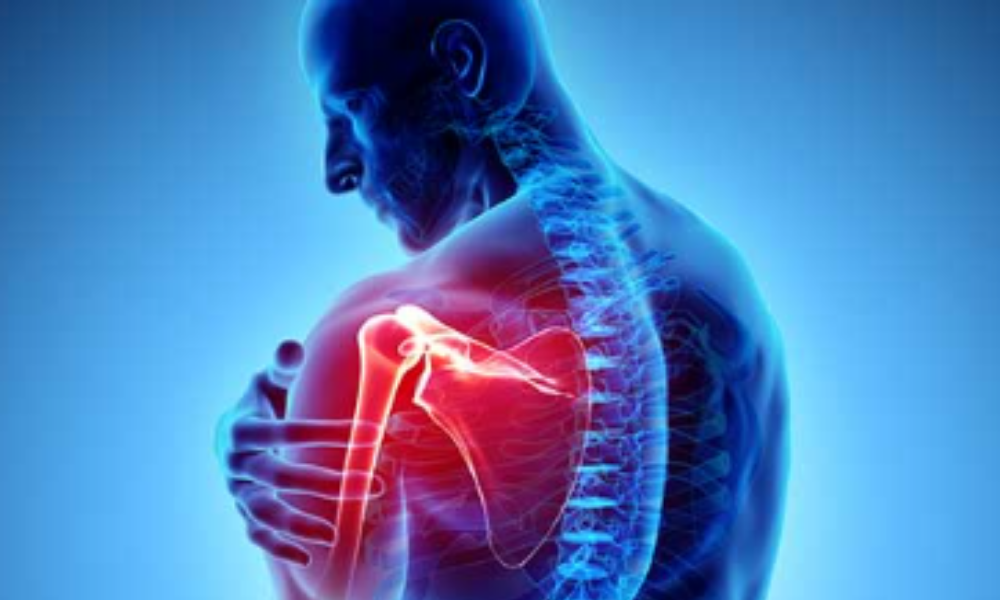
Shoulder pain is one of the commonest issues that I and other therapists see in practice. It’s often a secondary problem to lower and/or upper back pain.
The majority of people I see have been given a diagnosis of either impingement, frozen shoulder (also known as adhesive capsulitis), or a rotator cuff injury. Accurate assessment and possibly imaging is important for each of these problems. However, shoulder dysfunction can be multifaceted and swamped with compensations your body has put in place to try and protect your body. It can be a tricky area of the body to diagnose, treat and rehabilitate.
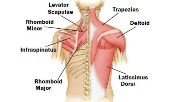
Image source Learn Anatome
1. Biomechanical dysfunction (sometimes referred to as scapular dyskinesis)
The shoulder is often called a complex rather than a joint because it relies on several joints to articulate. It effectively free-floats over the rib cage with stability and movement controlled by 17 muscles. The only connection to the main skeleton is via the collarbone, which articulates with the sternum.
The head of the upper arm (humerus) articulates with the scapula in a shallow joint called the glenohumeral joint. The shallow nature of the joint allows for a wide range of motion under normal alignment but this is at the expense of stability. To assist with stability there are four muscles that help pull the joint together. These are the rotator cuff muscles. In addition to stabilising the joint, they play a role in moving the arm but often only initiate movement. Larger muscles such as the deltoids, pecs, latissimus dorsi and trapezius add the strength and fuller range of motion. If any of these muscles are inhibited or overactive, then the full function of the shoulder and arm will be affected. Spinal alignment and stability is important for good shoulder range of motion. Try elevating your arm out to the side standing upright and then doing it again whilst slumping!
2. Biceps tendinopathy
Pain at the front of the shoulder can be a number of things, but one muscle that often gets overlooked in assessment is the biceps. The biceps muscle has two tendon attachments at its upper end into the scapula (shoulder blade). Both of these can be a cause of pain at the front of the shoulder but more commonly the long head of the biceps which sits in a groove at the head of the humerus.
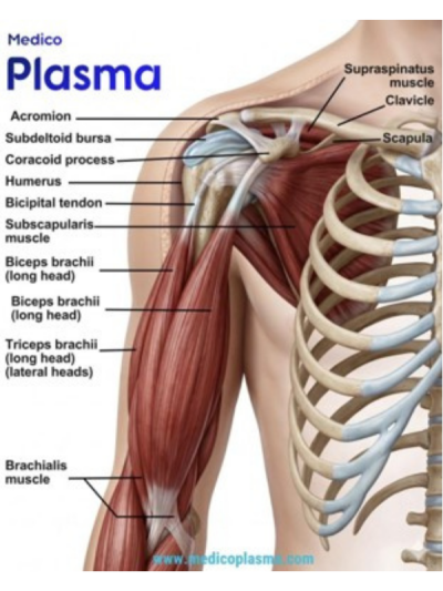
Image source Medico Plasma
Typical signs include pain with the arm outstretched in front, carrying heavy bags, and throwing actions. Posture plays an important role in the stability of the shoulder and biceps alignment. Rounded shoulders cause undue stress on the tendons and can cause inflammation and pain.
Remedial advice is to avoid heavy lifting, ice to reduce inflammation and posture awareness. Chronic pain may also require imaging to be carried out.
3. Rotator cuff injuries
There are four rotator cuff muscles (RCM) that serve to initiate movement and hold the shoulder joint together (humeral head to glenoid capsule).
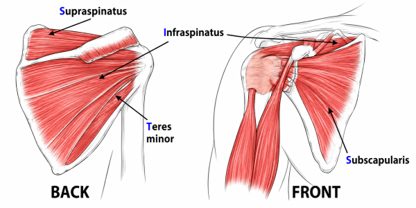
Image source Pixabay
RCM injuries can occur due to acute trauma injuries, typically when the shoulder joint is challenged by a sudden pulling, rotation or pushing movement. But they can also be due to slower micro-trauma due to factors such as age, poor posture and repetitive movements.
Typical signs – weakness, pain with movement or load-bearing, crepitus (clicking) with movement
Physiotherapy can help to rehabilitate these muscle injuries. If severe, surgery may be required.
An interesting fact about diagnosed rotator cuff injuries is that around 50% of minor tears are asymptomatic. Therefore, you could take a group of 100 people with no shoulder pain, ultrasound their shoulders and find some RCM pathology even though they have no pain. This means that the conclusion that the cause of pain is the RCM is often a misdiagnosis, especially where there has been no history of trauma.
4. Calcific tendonitis
This occurs when the rotator cuff tendons deposit calcium in the shoulder joint. It is most common with the supraspinatus muscle. It can occur due to degeneration (age or injury related), metabolic conditions or prolonged reduced blood flow.
The cycle of calcific tendonitis is two-fold – the calcification stage and the reabsorption phase. The latter is when the calcium deposits break down and generate more inflammation and pain.
Imaging such as ultrasound is the best diagnostic solution to establish this pathology.
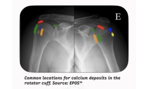
Image source EPOS
Typical signs include pain with elevation of the arm above the shoulder and weight-bearing.
5. Frozen Shoulder
Otherwise known as adhesive capsulitis, the causes of frozen shoulder are not clearly understood. Doctors know that it presents as a thickening and restriction of the joint capsule and that it is more common in women than men and usually occurs between the ages of 40 and 60.
It is more common to occur in the non-dominant arm. If you have had it in one shoulder, it is not uncommon to get it in the other, often years later.
There are usually 3 phases: (1) freezing; (2) frozen; (3) thawing. Each can last 6-12 months, or longer for thawing, making this condition more chronic than most.
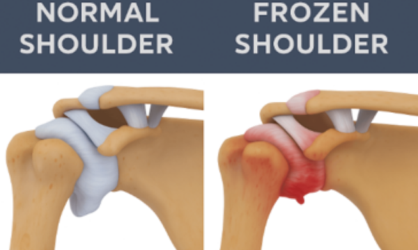
Image source Perth Shoulder Physio
Typical signs include pain at night, severe immobility and pain, lying on the affected shoulder, previous history in opposite arm. An interesting diagnostic feature of frozen shoulder is that passive motion (someone else moving the arm) is often more restrictive and painful than active motion (self-initiated).
Common predispositions include trauma, diabetes (both T1 and T2) and thyroid disorders.
It is usually very treatable with physiotherapy or other manual therapies but can take time to resolve.
In summary, Assessment of shoulder pain can be complex and should be carried out by a qualified professional. If you suffer from any of the above issues or other shoulder related issues, it’s best to seek advice from a medically trained specialist such as a medical doctor, physiotherapist, chiropractor or osteopath. Biomechanical shoulder pain is often attributed to problems elsewhere in the body because the alignment of the shoulder is closely affected by the alignment of the spine and the muscles that coordinate it.
Main – Image source JohnHopkinsUniversity
About the Author: Robert Spratley